
The anatomy of a long bone worksheet provides a comprehensive overview of the structure, function, and clinical significance of long bones. This detailed guide explores the various components of a long bone, including the diaphysis, epiphysis, metaphysis, periosteum, endosteum, and medullary cavity.
It also delves into the histology of long bones, examining the compact and spongy bone, Haversian and Volkmann’s canals, lacunae, and canaliculi.
Furthermore, the worksheet covers the blood supply and innervation of long bones, discussing the nutrient artery, nutrient foramen, epiphyseal arteries, periosteal arteries, venous drainage, and innervation. It also explores the growth and development of long bones, including endochondral ossification, the role of the growth plate, factors affecting bone growth, hormonal regulation, and stages of bone development.
Introduction
A long bone is a type of bone that is longer than it is wide. Long bones are found in the limbs, such as the arms and legs. They are responsible for providing support and movement to the body.
The purpose of this worksheet is to help you learn about the anatomy of a long bone. You will learn about the different parts of a long bone and their functions.
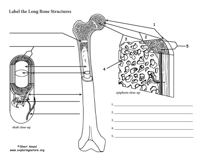
Parts of a Long Bone
- Diaphysis: The diaphysis is the main shaft of the bone. It is made up of compact bone, which is very dense and strong.
- Epiphysis: The epiphysis is the ends of the bone. It is made up of spongy bone, which is less dense than compact bone. The epiphysis is covered by a layer of cartilage, which helps to protect the bone and reduce friction.
- Metaphysis: The metaphysis is the region between the diaphysis and the epiphysis. It is made up of a mixture of compact and spongy bone.
- Articular cartilage: The articular cartilage is a layer of cartilage that covers the ends of the bone. It helps to reduce friction and wear and tear on the bone.
- Periosteum: The periosteum is a thin layer of tissue that covers the outside of the bone. It contains blood vessels and nerves that supply the bone with nutrients and oxygen.
- Endosteum: The endosteum is a thin layer of tissue that lines the inside of the bone. It contains blood vessels and nerves that supply the bone with nutrients and oxygen.
- Bone marrow: The bone marrow is a soft tissue that fills the inside of the bone. It produces red blood cells, white blood cells, and platelets.
Anatomy of a Long Bone
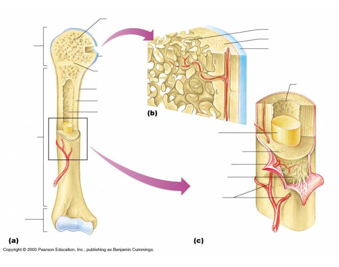
Diaphysis
The diaphysis is the main shaft of a long bone. It is responsible for providing structural support and protection to the bone. The diaphysis is made up of dense, compact bone tissue that is arranged in concentric layers. The outer layer of the diaphysis is called the cortex, which is thicker than the inner layer called the medulla.
Epiphysis
The epiphysis is the rounded end of a long bone. It is covered by a layer of cartilage called the articular cartilage, which allows the bone to articulate with other bones at the joints. The epiphysis is made up of cancellous bone tissue, which is less dense than compact bone tissue and contains a network of interconnected spaces filled with bone marrow.
Metaphysis
The metaphysis is the region of the bone that connects the diaphysis to the epiphysis. It is made up of a mixture of compact and cancellous bone tissue. The metaphysis is responsible for bone growth and development. During childhood, the metaphysis contains a growth plate, which is a layer of cartilage that allows the bone to grow in length.
Periosteum
The periosteum is a tough, fibrous membrane that covers the outer surface of the bone. It is responsible for providing nutrition to the bone, as well as for anchoring tendons and ligaments to the bone.
Endosteum
The endosteum is a thin membrane that lines the inner surface of the bone. It is responsible for lining the medullary cavity and for regulating the exchange of nutrients and waste products between the bone and the blood.
Medullary Cavity
The medullary cavity is the hollow space within the diaphysis of a long bone. It is filled with bone marrow, which is a soft tissue that produces blood cells and stores fat.
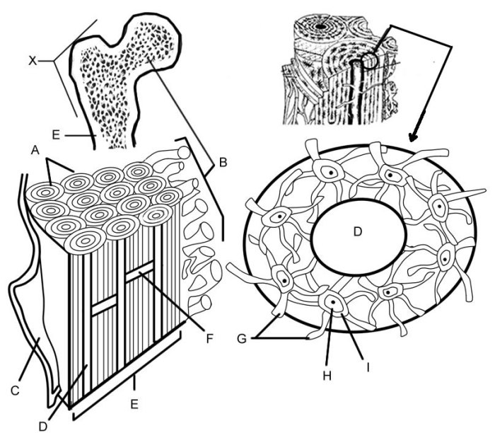
Histology of a Long Bone: Anatomy Of A Long Bone Worksheet

The histology of a long bone refers to the microscopic structure and organization of its tissues. It comprises two primary types of bone tissue: compact bone and spongy bone, each with distinct characteristics and functions.
Compact Bone
Compact bone forms the dense outer layer of a long bone, providing strength and rigidity. It is characterized by tightly packed Haversian systems, which are cylindrical units arranged parallel to the long axis of the bone.
Haversian Canals
Haversian canals are central channels within each Haversian system. They contain blood vessels, nerves, and lymphatic vessels, supplying nutrients and oxygen to the bone cells.
Lacunae and Canaliculi, Anatomy of a long bone worksheet
Lacunae are small cavities within the bone matrix that house osteocytes, the mature bone cells. Canaliculi are tiny channels that radiate from the lacunae, allowing nutrients and waste products to diffuse between the osteocytes and the Haversian canals.
Spongy Bone
Spongy bone, also known as cancellous bone, forms the inner core of a long bone. It is composed of a network of thin, interconnected trabeculae, which are bony struts that provide structural support while allowing for weight reduction.
Volkmann’s Canals
Volkmann’s canals are larger channels that traverse the spongy bone, perpendicular to the Haversian canals. They connect the Haversian systems and allow for the passage of blood vessels and nerves into the inner bone.
Blood Supply and Innervation of a Long Bone
Long bones have a complex blood supply and innervation that supports their growth, repair, and function. The nutrient artery is the primary blood supply to the bone, entering through the nutrient foramen. Epiphyseal arteries supply the ends of the bone, while periosteal arteries supply the outer surface.
Venous drainage occurs through a network of veins that empty into the systemic circulation. Innervation is provided by nerves that enter the bone through nutrient foramina and periosteal foramina.
Nutrient Artery and Foramen
The nutrient artery is a large artery that enters the bone through the nutrient foramen, which is located on the diaphysis. The nutrient artery branches into ascending and descending branches that supply the bone marrow and the inner layers of the bone.
Epiphyseal Arteries
Epiphyseal arteries supply the ends of the bone, the epiphyses. These arteries enter the epiphyses through foramina in the articular cartilage. Epiphyseal arteries are important for bone growth and repair.
Periosteal Arteries
Periosteal arteries supply the outer surface of the bone, the periosteum. These arteries enter the periosteum through small foramina in the bone. Periosteal arteries are important for bone growth and repair.
Venous Drainage
Venous drainage of a long bone occurs through a network of veins that empty into the systemic circulation. The veins are located in the bone marrow, the periosteum, and the surrounding soft tissues.
Innervation
Innervation of a long bone is provided by nerves that enter the bone through nutrient foramina and periosteal foramina. The nerves supply the bone marrow, the periosteum, and the surrounding soft tissues.
Growth and Development of a Long Bone
Long bone growth is a complex process involving endochondral ossification, the formation of bone from cartilage. The growth plate, a specialized region at the ends of long bones, plays a crucial role in this process.
Endochondral Ossification
Endochondral ossification involves the replacement of cartilage with bone. It occurs in the following stages:
- Chondrocytes(cartilage cells) proliferate and form a cartilage model of the bone.
- Blood vessels invade the cartilage, bringing nutrients and oxygen.
- Osteoblasts(bone-forming cells) deposit bone matrix around the cartilage.
- The cartilage is gradually resorbed by chondroclasts(bone-resorbing cells), leaving behind a cavity that is filled with bone.
Role of the Growth Plate
The growth plate is a layer of cartilage located at the ends of long bones. It is responsible for longitudinal bone growth. As the chondrocytes in the growth plate proliferate and differentiate, new cartilage is formed, which is then replaced by bone through endochondral ossification.
Factors Affecting Bone Growth
Several factors can influence bone growth, including:
- Genetics:Genes play a significant role in determining bone size and shape.
- Nutrition:Adequate intake of calcium, vitamin D, and other nutrients is essential for bone growth.
- Physical activity:Regular exercise can stimulate bone growth.
- Hormones:Growth hormone, parathyroid hormone, and thyroid hormone are among the hormones that regulate bone growth.
Hormonal Regulation of Bone Growth
Growth hormone is the primary hormone responsible for longitudinal bone growth. It stimulates the proliferation of chondrocytes in the growth plate. Other hormones, such as parathyroid hormone and thyroid hormone, also play a role in bone growth.
Stages of Bone Development
Bone development occurs in several stages:
- Embryonic stage:The skeletal system begins to develop during embryonic development.
- Fetal stage:Bones begin to ossify, replacing cartilage with bone.
- Childhood:Bone growth continues rapidly, reaching its peak during puberty.
- Adolescence:Bone growth slows down and eventually stops.
- Adulthood:Bone remodeling continues to maintain bone strength and integrity.
Clinical Significance

Long bones play a vital role in maintaining the structural integrity of the body and facilitating movement. However, they are also susceptible to various injuries and disorders that can have significant clinical implications.
Fractures
Fractures are breaks or cracks in the bone, often resulting from trauma or excessive force. They can range from minor hairline fractures to severe, life-threatening injuries. Fractures can cause pain, swelling, bruising, and impaired mobility. Treatment typically involves immobilization, pain management, and, in some cases, surgery.
Metabolic Disorders
Long bones are involved in calcium and phosphorus metabolism. Disorders such as osteoporosis and Paget’s disease can affect bone density and structure, making them more susceptible to fractures. Osteoporosis is characterized by decreased bone density, while Paget’s disease involves abnormal bone remodeling, leading to enlarged and weakened bones.
Mobility Impairment
Injuries to long bones can significantly impact mobility. Fractures, dislocations, and other injuries can impair movement, range of motion, and coordination. Rehabilitation is crucial for restoring mobility and preventing long-term complications.
Forensic Investigations
Long bones play a crucial role in forensic investigations. The examination of bone morphology, density, and other characteristics can help determine the age, sex, and stature of an individual. Bone analysis can also provide insights into the cause and manner of death, particularly in cases of trauma.
Medical Research
Research on long bones has led to advancements in understanding bone biology, injury management, and disease prevention. Studies on bone regeneration, tissue engineering, and biomechanics have contributed to the development of new treatments and technologies for bone-related conditions.
Popular Questions
What is the purpose of the periosteum?
The periosteum is a fibrous membrane that covers the outer surface of long bones and plays a crucial role in bone growth, repair, and nutrition.
What is the significance of the medullary cavity?
The medullary cavity is a hollow space within the diaphysis of long bones that contains bone marrow, which produces red and white blood cells.
How does endochondral ossification contribute to bone growth?
Endochondral ossification is a process by which cartilage is replaced by bone, contributing to the growth and development of long bones.
